
About Xe MRI
Imaging with hyperpolarized noble gases, such as xenon, is an emerging technique that provides researchers and clinicians with a novel, noninvasive way to assess lung health. Hyperpolarized xenon imaging—commonly referred to as ¹²⁹Xe MRI—offers highly sensitive, detailed regional measurements of lung structure and function that are not achievable with conventional imaging methods.
Using a specialized device called a polarizer, xenon gas is hyperpolarized, making it detectable by MRI. During the procedure, patients inhale the hyperpolarized gas from a collection bag, similar to breathing in helium from a balloon. While the patient briefly holds their breath, MRI images are acquired.
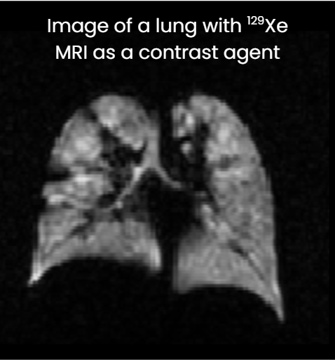
In this process, ¹²⁹Xe acts as a contrast agent, producing a clear, “illuminated” view of the lungs. This allows clinicians and researchers to evaluate regional ventilation, gas exchange, and microstructure with exceptional detail. As a result, Xe MRI is particularly valuable for detecting early functional changes in a wide range of respiratory diseases.
Now FDA approved, Xe MRI has become an essential tool in academic research and clinical trials. It has contributed to important advances in understanding lung disease characteristics and progression, and it is increasingly used to inform clinical decision-making for patients ages 6 and up. In addition, ¹²⁹Xe MRI is now at the forefront of clinical trial endpoints, serving as a powerful tool for evaluating the effectiveness of emerging therapies in pharmaceutical and biomedical research.
The Xe MRI Clinical Trials Consortium is proud to have helped pioneer this technology and advance its use as a critical resource for improving lung health.
What is Xe MRI?
Xe MRI: What to Expect
The video below, courtesy of Cincinnati Children’s, provides a helpful overview of what patients can expect when undergoing a hyperpolarized xenon MRI scan.